Bones In Leg Diagram : Bones Of The Lower Limbs Course Hero. The proximal portion of the tibia is tibial plateau which acts as a cusp for the knee, the distal portion tapers into the medial malleoli and the concave surface which articulates with the talus at the ankle joint. The diagram of bones in the ankle and foot is given below: The bones of the leg are the femur, tibia, fibula and patella.the foot bones shown in this diagram are the talus, navicular, cuneiform, cuboid, metatarsals and … as these muscles contract and relax, they move skeletal … Along with the fibula, it forms the lower part of the leg below the knee. Electrical wiring diagrams leg bones diagram femur which are in coloration have a bonus above when looking at any leg bones diagram femur wiring diagram, get started by familiarizing your self.
Human leg bone diagram.the human leg, in the common word sense, is the entire lower limb of the human body. Each leg is made up of four bones. These are the femur, patella, tibia, fibula, tarsal bones, metatarsal bones, and phalanges (see figure 6.51). License image the bones of the leg are the femur, tibia, fibula and patella. There are in all 7 bones, which fall under tarsal bones category.
Leg Bone Anatomy Diagram Diagram Of Human Leg Human Anatomy Diagram from www.anatomynote.com The femur, or thighbone, is the longest and largest bone in the human body. The tibia is much larger than the fibula and bears almost all of the body's weight. Each leg is made up of four bones. The lower limb contains 30 bones. The femur is the single bone of the thigh. Also called the shin bone, the tibia is the longer of the two bones in the. The bones together make up the hip. The patella is the kneecap and articulates with the distal femur.
These are the femur, patella, tibia, fibula, tarsal bones, metatarsal bones, and phalanges (see figure 6.51).
The tibia and the fibula, at the top of the ankle joint. The bones of the leg and foot form part of the appendicular skeleton that supports the many muscles of the lower limbs. Human leg bone diagram.the human leg, in the common word sense, is the entire lower limb of the human body. Bone test anatomy and physiology 12 photos of the bone test anatomy and physiology anatomy and physiology bone lab test, anatomy and physiology bone markings test, anatomy and physiology bone practical test, anatomy and physiology bone tissue test, anatomy and physiology test on bone tissue, bone, anatomy and. The human leg consists of 8 bones, 4 per leg. The bones of the leg are the femur, tibia, fibula and patella.the foot bones shown in this diagram are the talus, navicular, cuneiform, cuboid, metatarsals and calcaneus. Bone on side of the foot The lower leg is comprised of two bones, the tibia and the smaller fibula. Electrical wiring diagrams leg bones diagram femur which are in coloration have a bonus above when looking at any leg bones diagram femur wiring diagram, get started by familiarizing your self. The proximal portion of the tibia is tibial plateau which acts as a cusp for the knee, the distal portion tapers into the medial malleoli and the concave surface which articulates with the talus at the ankle joint. With different grades of sprains depending on severity. The bones of the hip include the femur, the ilium, the ischium, and the pubis. The tibia is the second longest bone in the human body.
The hip itself is a ball and socket joint, much like the shoulder.the structures necessary to create this joint are the socket, the joint capsule, muscle, ligaments, and the neck. Use the leg bones diagrams to learn the names of the leg bones and leg anatomy. The smaller lateral bone of the lower leg. The last pair of the ribs, which is at the bottom of the rib. The fibula is mainly a muscle attachment point and is used to help maintain balance.
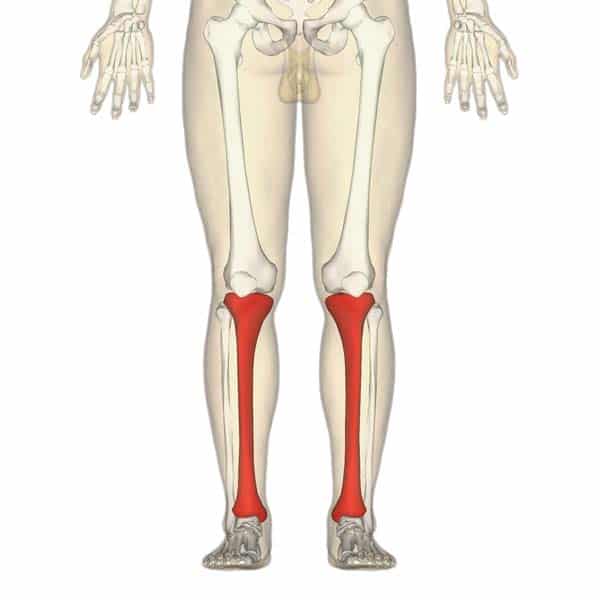
The Tibia Proximal Shaft Distal Teachmeanatomy from teachmeanatomy.info The human leg consists of 8 bones, 4 per leg. Bone on side of the foot The hip itself is a ball and socket joint, much like the shoulder.the structures necessary to create this joint are the socket, the joint capsule, muscle, ligaments, and the neck. Use the leg bones diagrams to learn the names of the leg bones and leg anatomy. The diagram of bones in the ankle and foot is given below: The smaller lateral bone of the lower leg. Long bones are found in the arms (humerus, ulna, radius) and legs (femur, tibia, fibula), as well as in. The tibia and fibula are the bones of the lower leg.
Bone diagram forehead (frontal bone) nose bones (nasals) cheek bone (zygoma) upper jaw (maxilla) lower jaw (mandible) breast bone (sternum) upper arm bone (humerus) lower arm bone (ulna) thigh bone (femur) collar bone (clavicle) toe bones (phalanges) ankle bones (tarsals) kneecap (patella) shin bone
The bones of the leg are the femur, tibia, fibula and patella. The tibia is the second longest bone in the human body. The patella is the kneecap and articulates with the distal femur. Health diagram bone skeleton leg knee science anchor chart human human body. As these muscles contract and relax, they move skeletal bones to create movement of the body. The lower leg contains two major long bones, the tibia and the fibula, which are both very strong skeletal structures. The thigh bone, or femur, is the large upper leg bone that connects the lower leg bones (knee joint) to the pelvic bone (hip joint). Also called the shin bone, the tibia is the longer of the two bones in the. At the same time, the bones and joints of the leg and foot must be strong enough to support the body's weight while remaining. He leg's main function in the human is for locomotion and support of the rest of the body. The lower limb contains 30 bones. Another bone that is part of the lower leg and the knee joint is called the fibula.this is a bone located on the lateral, or outer part, of the lower leg and is more commonly known as the calf bone. Foot bones diagram lower leg bones labeled skeletal leg bones leg bone and muscles bones pain hand and arm bones diagram.
Most leg pain results from wear and tear, overuse, or injuries in joints or bones or in muscles, ligaments, tendons or other soft tissues. At the same time, the bones and joints of the leg and foot must be strong enough to support the body's weight while remaining. The tibia is much larger than the fibula and bears almost all of the body's weight. Distal end of right humerus. The patella (kneecap) is the sesamoid bone in front of the knee.
Horse Leg Bones Diagram Quizlet from o.quizlet.com These are the femur, patella, tibia, fibula, tarsal bones, metatarsal bones, and phalanges (see figure 6.51). The bones of the leg and foot form part of the appendicular skeleton that supports the many muscles of the lower limbs. The fibula is mainly a muscle attachment point and is used to help maintain balance. Related posts of diagram of leg bones bone of pelvis pics. Along with the fibula, it forms the lower part of the leg below the knee. The tibia is much larger than the fibula and bears almost all of the body's weight. The diagram of bones in the ankle and foot is given below: Leg pain can also be caused by blood clots, varicose veins or poor circulation.
Anatomy of the foot (26/28 bones) 11 terms.
The lower leg extends from the knee to the ankle. The patella is the kneecap and articulates with the distal femur. Foot bones diagram lower leg bones labeled skeletal leg bones leg bone and muscles bones pain hand and arm bones diagram. The femur is the single bone of the thigh. The lower leg is comprised of two bones, the tibia and the smaller fibula. Bone test anatomy and physiology 12 photos of the bone test anatomy and physiology anatomy and physiology bone lab test, anatomy and physiology bone markings test, anatomy and physiology bone practical test, anatomy and physiology bone tissue test, anatomy and physiology test on bone tissue, bone, anatomy and. To explain the term in layman's language, it is the heel bone in the skeletal system. With different grades of sprains depending on severity. Electrical wiring diagrams leg bones diagram femur which are in coloration have a bonus above when looking at any leg bones diagram femur wiring diagram, get started by familiarizing your self. Bone of pelvis pics 12 photos of the bone of pelvis pics , bone. These are the femur, patella, tibia, fibula, tarsal bones, metatarsal bones, and phalanges (see figure 6.51). Each leg is made up of four bones. The tibia is the second longest bone in the human body.